Diabetic Retinopathy by Dr Tara Mary George
What Is Diabetic Retinopathy?
Diabetic retinopathy is a condition where the fine blood vessels or capillaries that supply oxygen and nutrients to the retina get damaged due to high levels of blood sugar in the blood seen in diabetes mellitus.
How Else Can Diabetes Affect The Eye?
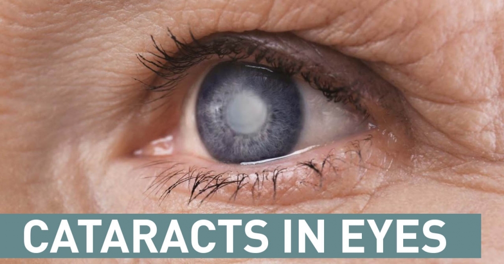
Diabetes can alter the lens clarity and diabetics develop cataracts at an earlier age with a higher rate compared to the normal population. Diabetics are more prone to dry eyes, eyelid infections and glaucoma. Other retinal blood vessel disorders such as retinal vein and artery occlusion (stroke in the eye) also occur more commonly in diabetics.
What Are The Types/ Stages of Diabetic Retinopathy?
- Non-proliferative retinopathy. It can be divided into mild, moderate, and severe forms. The mild and moderate forms may reverse or remain stable with good control of Diabetes Mellitus
- Proliferative retinopathy
What Are The Symptoms of Diabetic Retinopathy?
- Early-stage – NO SYMPTOMS
- Gradual or sudden blurring of vision
- ‘Floaters’- blackspots / web-like spots in the visual field

- Problems in reading books or signage
- Distortion of the straight line

Are you experiencing any symptoms mentioned above? Don’t hesitate to contact our eye centre and make an appointment today.
Risk Factors of Diabetic Retinopathy
- People with a duration of diabetes more than 10 years
- Poorly controlled diabetics
- Those with other organ damage such as kidney damage (nephropathy) as the eyes and kidneys share similar blood vessels
- Smokers
- Pregnant diabetics
- Diabetics with anemia
How to reduce risk of visual loss?
- Commit to managing blood glucose
- Keep your blood pressure and cholesterol under control
- Ensure yearly eye examination
- Stop smoking
- Pay attention to vision changes
What will happen during the eye check-up?
Eye check up is the first thing we are going to do for diabetic retinopathy treatment. Visual acuity and eye pressure will be checked. Retinal photography with special cameras and the photographs are graded.
These examinations do not blur the vision. If required, dilated retinal examination will be done by an ophthalmologist. The dilated pupils may blur your vision slightly for up to 2 hours post examination.
Back